Free Radiology Technician Course (1year Diploma Course)
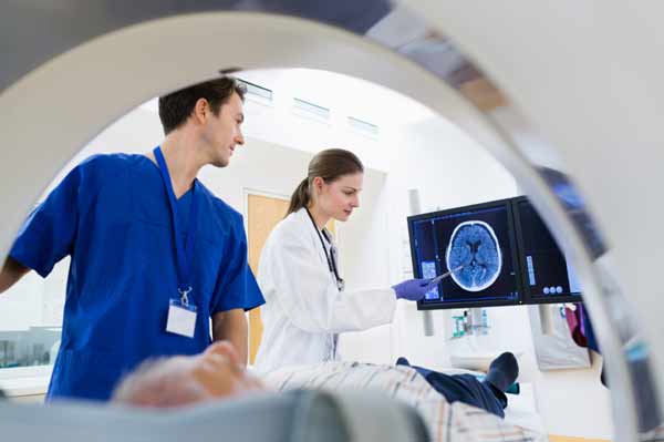
Brief Job Description:- Radiology Technicians perform diagnostic imaging examinations such as X-rays, CT and MRI scans under the guidance of a Radiologist. Radiology Technicians are responsible for preparing patients and operating equipment for the test/tests, besides keeping patient records, adjusting equipment’s based on patient need and test recommended and maintaining equipment.
Personal Attributes:- Radiology Technicians must be able to interact with patient and their attendants and be a team players. They must also be polite and be able to calm and placate upset patients (and accompanying members). They should be able to work for long period of time in standing position and must be able direct, transfer, help patients reach the test location Radiology Technician Course

Processing radiographic images, Carrying out quality control tests on images obtained Rediology Technician Course:-
To be competent, the user/individual on the job must be able to:
PC1. Explain the principles ofradiographic imaging
PC2. Apply knowledge ofradiographic imaging to the production ofradiographs and the assessment of image quality
PC3. Understand the construction and operation of image processing equipment
PC4. Control andmanipulate parameters associated with exposure and processing to produce a required image of desirable quality
PC5. Perform X‐ray film /image processing techniques(including dark room techniques)
PC6. Explain and implement the fundamentals, concepts and applications of processing of imagesin digital form using computer based systems
PC7. Carry out quality control for automatic film processing, evaluate and act on
Taking the advice of a radiologist on the scans performed , Documenting diagnosis and comments of tTaking the advice of a radiologist on the scans performed , Documenting diagnosis and comments of the
Taking the advice of a radiologist on the scans performed ,Documenting diagnosis and comments of the radiologist in a report for the patient:- To be competent, the user/individual on the job must be able to:
PC1. Correctly identify anatomical features on the radiographs and identity some major pathological and traumatic conditions
PC2. Seek the advice ofthe Radiologist on conditionsidentified
PC3. Document the comments and diagnosis of the Radiologist in a report for the patient
PC4. Explain the diagnosis and comments and the report to the patient if required


Taking precautionary measures to avoid the reactions, Recognising the contrast induced reaction Rediology Technician Course:- To be competent, the user/individual on the job must:
PC1. Know the patient’s medical history
PC2. Select proper agent to be used PC3. Promptly recognize and assess the
PC4. Ensure immediate availability of necessary equipment and drugsin case of reaction
PC5. Know the correct medications and other treatment options
Free Radiology Technician Course (1year Diploma Course)
PC6. Know the different types of adverse reactions Rediology Technician Course.
PC7. Recognise the contraindications of allergic reactions
Communicating with individuals, patients, theirfamily and others about health issues:-

To be competent, the user/individual on the job must be able to:
PC1. Respond to queries and information needs of all individuals
PC2. Communicate effectively with all individualsregardless of age, caste, gender, community or other characteristics
PC3. Communicate with individuals at a pace and level fitting their understanding, without using terminology unfamiliarto them
PC4. Utilise all training and information at one’s disposal to provide relevant information to the individual
PC5. Confirm that the needs ofthe individual have been met
PC6. Adhere to guidelines provided by one’s organisation orregulatory body relating toconfidentiality
PC7. Respect the individual’s need for privacy PC8. Maintain any recordsrequired at the end of the interaction
Core Skills/ Generic Skills:- The user/individual on the job needsto know and understand how to:
SA1. Write short notesto co‐workers and clericalstaff to compile information about particular patients, describe unusual pathologies or ask for on‐site reference material

Free Radiology Technician Course (1year Diploma Course)
SA2. Write brief observations about pathologiesthat may affect diagnoses on patients’charts
SA3. Write detailed notes aboutscans done Rediology Technician Course.
SA4. Write descriptions of accidents and incidents on reporting forms when something unusual occurs during patient exams orscanning procedures
SA5. Write memosto advise, inform or directstaff working in other hospital or clinic departments or units
SA6. Complete patients’ medical history forms by entering the patients’ names, treatmentsreceived to date and current medical conditions


Reading Skills:- The user/individual on the job needsto know and understand how to:
SA7. Read scan instructionsin notes attached to patients’ files
SA8. Read communications aboutscheduling, training and updatesto internal proceduresfromco‐workers,supervisors or hospital administrators
SA9. Read protocol updates and hospital policy changes
SA10. Read and follow allspecified proceduresin the multi‐page treatment prescriptions prepared by referring physicians
SA11. Review protocolsforscanning and identifying non‐routine or atypical pathologiesinprocedure manuals
SA12. Read reports of varying lengths completed by physicians, hospital or clinic administratorsandsupervising technologists
SA13. Read user manualsfor varioustypes ofradiological equipment when troubleshooting faults with scanners orimaging computers
Oral Communication (Listening and Speaking skills) Rediology Technician Course:- The user/individual on the job needsto know and understand how to:
SA14. Speak to patientsto explain protocolsfor procedures or examinations, obtain information aboutthe patient’sstatus and discuss current diagnoses and treatmentoptions
SA15. Speak with reception and clericalstaff to determine and confirm the number of appointmentsforthe day, request patient information from files an loggings of appointmentsfor patientsrequiring additionaltesting ortreatment
SA16.Discussscheduling,treatmentroomassignments andworkload responsibilities with employees and co‐workers Rediology Technician Course.
SA17.Ordersuppliessuch as contrast media and radioactive pharmaceuticalsfrom suppliers and hospital dispensaries
SA18.Discuss proceduralsuggestions, equipmentmalfunctions and personnel problems with the seniortechnologists, unit or departmentsupervisors or administrative staff
SA19. Comfort patients who may be frightened or upset during scanning procedures
SA20. Discuss patients’status with nurses,social workers, dieticians or other members ofthe extended health care team
Professional Skills:- The user/individual on the job needsto know and understand how to:
SB1. Choose the correct film size for the sizes of the areasto be scanned
SB2. Decide on a course of action when physicians have requested types of radiographs orscansfor patients who cannot be positioned in a typical way
SB3. Decide which patients will be processed first when they receive multiple requisitions at the same time, or during emergencies
SB4. Decide if examinations can be completed under contraindicative or complicating circumstances
Plan and Organize:- The user/individual on the job needsto know and understand:
SB5. How to determine the order and priority of work taskssubject to confirmation or approval fromsupervisors
SB6. How to integrate work plans with those of the extended health care teams Rediology Technician Course.
SB7. How to schedule daily work priorities based on the demands of the clinic, laboratory or hospital SB8. How to schedule patient‐load based on emergency or appointment priority
Customer Centricity:- The user/individual on the job needsto know and understand how to:
SB9. Comfort patients who may be frightened or upset during scanning procedures
Free Radiology Technician Course (1year Diploma Course)
SB10. Liaise with members of the extended health care team to ensure the needs of the patient are taken care of
Problem Solving:- The user/individual on the job needsto know and understand how to:
SB11. Indicate importantscanning parameters on x‐rays orscanned images,such as appropriate spatial or directional indicators when these have been neglected earlierin the process
. Recommend alternate scan types/ positions and discuss these with the radiologist when the scan recommended by the physician is not possible or is difficult forthe patient
SB13. Re‐schedule appointments when patients arriving for exams are late or have not taken the necessary pre‐appointment measuressuch asfasting orrefraining from takinginterferingmedications
SB14. Troubleshootradiological equipment when aminorfault occurs

Analytical Thinking Radiology Technician Course:- The user/individual on the job needs to know and understand how to:

SB15. Analyse the prescription ofthe patient and decide on the best position to take the recommended scan
SB16. Analyse the scan imagesto determine quality and clarity
SB17. Analyse the inventory ofsuppliesto decide when to place an order toreplenishthese
CriticalThinking:- The user/individual on the job needsto know and understand how to:
SB18. Make preliminary judgements about the seriousness of patients’ injuries
SB19. Evaluate the quality ofradiographs, digital images and scans Rediology Technician Course.
Guidelines for Assessment:-
- Criteria for assessment for each Qualification Pack will be created by the Sector Skill Council. Each Performance Criteria (PC) will be assigned marks proportional to its importance in NOS. SSC will also lay down proportion of marks for Theory and Skills Practical for each
PC 2. The assessment for the theory part will be based on knowledge bank of questions created by the
SSC 3. Individual assessment agencies will create unique question papers for theory part for each candidate at each examination/training center (as per assessment criteria below)
4. Individual assessment agencies will create unique evaluations for skill practical for every student at each examination/training center based on this criteria
5. To pass the Qualification Pack, every trainee should score as per assessment grid Rediology Technician Course.
6. In case of successfully passing only certain number of NOS’s, the trainee is eligible to take subsequent assessment on the balance NOS’s to pass the Qualification Pack
Follow radiological diagnostic needs of patients Rediology Technician Course:-
PC1. Explain the subdivisions of anatomy, terms of location and position, fundamental planes, vertebrate structure of man, organisation of the body cells and tissues
PC2. Explain the pathology of various systems: cardiovascular system, respiratory system, central nervous system, musculoskeletal system, GIT, GUT and reproductive system
PC3. Explain the pathology of radiation injury and malignancies
PC4. Understand specific requests of physicians with respect to the scans required
PC5. Take medical history of the patient and document it as required
PC6. Understand and interpret instructions and requirements documented by the physician in the patient’s prescription
PC7. Determine the radiological diagnostic tests required for the patient based on the physician’s prescription and the medical history

Prepare the patient and the room for the procedure:-
PC1. Prepare the room, apparatus and instruments for an x-ray, CT scan or MRI scan
PC2. Set up the X-ray machine, MRI machine or CT scan machine for the procedure
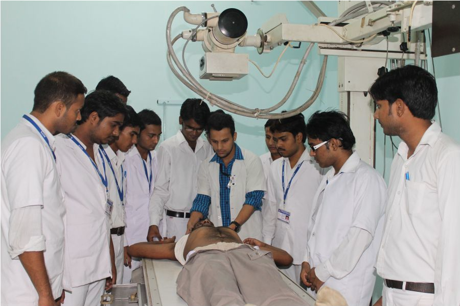
PC3. Position the patient correctly for an x-ray in the following positions:
a. Erect
b. Sitting
c. Supined
d. Prone
e. Lateral
f. Oblique
g. Decubitus
PC4. Explain relative positions of x-ray tube and patient and the relevant exposure factors related to these
PC5. Explain the use of accessories such as Radiographic cones, grid and positioning aids
PC6. Explain the anatomic and physiological basis of the procedure to be undertaken Rediology Technician Course.
PC7. Explain the radiographic appearances of both normal and common abnormal conditions where elementary knowledge of the pathology involved would ensure application of the appropriate radiographic technique
PC8. Position the patient correctly for a Computed Tomography scan
PC9. Position the patient correctly for an MRI scan
PC10. Apply modifications in positioning technique for various disabilities and types of subject
PC11. Explain the use of contrast materials for a CT scan and how to administer them under supervision of a radiologist
PC12. Explain the use of MRI Contrast agents and how to administer them under supervision of a radiologist
PC13. Manage a patient with contrast reaction
PC14. Explain the principles of radiation physics detection and measurement
PC15. Explain the biological effects of radiation
PC16. Explain the principles of radiation protection:
a. Maximum permissible exposure concept
b. Annual dose equivalent limits (ADEL) ALARA concept
c. International recommendations and current code of practice for the protection of persons
against ionising radiation from medical and dental use
PC17. Explain the use of protective materials:
a. Lead
b. Lead – impregnated substances
c. Building materials
d. Concept of barriers
e. Lead equivalents and variations
f. Design of x-ray tubes related to protection
. g. Structural shielding design (work-load, use factor, occupancy factor, distance Rediology Technician Course.
PC18. Explain the instruments of radiation protection, use of gonad shield and practical methods for reducing radiation dose to the patient
PC19. Ensure protection of self, patients, departmental staff and public from radiation through use of protection instruments and monitoring personnel and the work area
Operate and oversee operation of radiologic equipment:-
PC1. Describe the construction and operation of general radiographic equipment
PC2. Describe the construction and operation of advanced imaging equipment including CT and MRI
PC3. Reliably perform all non-contrast plain Radiography, conventional contrast studies and non-contrast plain radiography in special situations
PC4. Apply quality control procedures for all radiologic equipment
PC5. Control and manipulate parameters associated with exposure and processing to produce a required image of desirable quality
PC6. Practise the procedures employed in producing a radiographic image
PC7. Describe methods of measuring exposure and doses of radiographic beams
PC8. Help in administration of correct contrast dosage Rediology Technician Course.
PC9. Discuss and apply radiation protection principles and codes of practice
PC10. Demonstrate an understanding of processing of images in digital form and be familiar with recent advances in imaging
PC11. Set up the X-ray machine, MRI machine or CT scan machine for the procedure
PC12. Carry out routine procedures associated with maintenance of imaging and processing systems
PC13. Ensure protection of patients, departmental staff and public from radiation through use of protection instruments and monitoring personnel and the work area
Process radiographic images:-
PC1. Explain the principles of radiographic imaging
PC2. Apply knowledge of radiographic imaging to the production of radiographs and the assessment of image quality
PC3. Understand the construction and operation of image processing equipment
PC4. Control and manipulate parameters associated with exposure and processing to produce a required image of desirable quality
PC5. Perform X-ray film / image processing techniques (including dark room techniques)
PC6. Explain and implement the fundamentals, concepts and applications of processing of images in digital form using computer based systems
PC7. Carry out quality control for automatic film processing, evaluate and act on results
Prepare and document reports:-
PC1. Correctly identify anatomical features on the radiographs and identity some major pathological and traumatic conditions
PC2. Seek the advice of the Radiologist on conditions identified
PC3. Document the comments and diagnosis of the Radiologist in a report for the patient
Recognise contrast induced adverse reactions:-
PC1. Know the patient’s medical history
PC2. Select proper agent to be used
PC3. Promptly recognise and assess the reactions
PC4. Ensure immediate availability of necessary equipment and drugs in case of reaction
PC5. Know the correct medications and other treatment options
PC6. Know the different types of adverse reactions
PC7. Recognise the contraindications of allergic reaction
(infection control policies and procedures):-
PC1. Preform the standard precautions to prevent the spread of infection in accordance with organisation requirements
PC2. Preform the additional precautions when standard precautions alone may not be sufficient to prevent transmission of infection
PC3. Minimise contamination of materials, equipment and instruments by aerosols and splatter
PC4. Identify infection risks and implement an appropriate response within own role and responsibility
PC5. Document and report activities and tasks that put patients and/or other workers at risk
PC6. Respond appropriately to situations that pose an infection risk in accordance with the policies and procedures of the organization
PC7. Follow procedures for risk control and risk containment for specific risks
PC8. Follow protocols for care following exposure to blood or other body fluids as required
PC9. Place appropriate signs when and where appropriate
PC10. Remove spills in accordance with the policies and procedures of the organization
PC11. Maintain hand hygiene by washing hands before and after patient contact and/or after any activity likely to cause contamination
PC12. Follow hand washing procedures
PC13. Implement hand care procedures
PC14. Cover cuts and abrasions with water-proof dressings and change as necessary
PC15. Wear personal protective clothing and equipment that complies with Indian Standards, and is appropriate for the intended use
PC16. Change protective clothing and gowns/aprons daily, more frequently if soiled and where appropriate, after each patient contact
PC17. Demarcate and maintain clean and contaminated zones in all aspects of health care work
PC18. Confine records, materials and medicaments to a well-designated clean zone
PC19. Confine contaminated instruments and equipment to a well-designated contaminated zone
PC20. Wear appropriate personal protective clothing and equipment in accordance with occupational health and safety policies and procedures when handling waste
PC21. Separate waste at the point where it has been generated and dispose of into waste containers that are colour coded and identified
PC22. Store clinical or related waste in an area that is accessible only to authorised persons
PC23. Handle, package, label, store, transport and dispose of waste appropriately to minimise potential for contact with the waste and to reduce the risk to the environment from accidental release
PC24. Dispose of waste safely in accordance with policies and procedures of the organisation and legislative requirements
PC25. Wear personal protective clothing and equipment during cleaning procedures
PC26. Remove all dust, dirt and physical debris from work surfaces
PC28. Decontaminate equipment requiring special processing in accordance with quality management systems to ensure full compliance with cleaning, disinfection and sterilisation protocols
PC29. Dry all work surfaces before and after use
PC30. Replace surface covers where applicable
PC31. Maintain and store cleaning equipment
(within the limits of one’s competence andauthority):-
PC1. Adhere to legislation, protocols and guidelines relevant to one’s role and field of practice
PC2. Work within organisational systems and requirements as appropriate to one’s role
PC3. Recognise the boundary of one’s role and responsibility and seek supervision when situations are beyond one’s competence and authority
PC4. Maintain competence within one’s role and field of practice
PC5. Use relevant research based protocols and guidelines as evidence to inform one’s practice
PC6. Promote and demonstrate good practice as an individual and as a team member at all times
PC7. Identify and manage potential and actual risks to the quality and safety of practice
PC8. Evaluate and reflect on the quality of one’s work and make continuing improvements
(Ensure availability of medical and diagnostic supplies):-
PC1. Maintain adequate supplies of medical and diagnostic supplies
PC2. Arrive at actual demand as accurately as possible
PC3. Anticipate future demand based on internal, external and other contributing factors as accurately as possible
PC4. Handle situations of stock-outs or unavailability of stocks without compromising health needs of patients/ individuals
(Collate and Communicate Health Information):-
PC1. Respond to queries and information needs of all individuals
PC2. Communicate effectively with all individuals regardless of age, caste, gender, community or other characteristics
PC3. Communicate with individuals at a pace and level fitting their understanding, without using terminology unfamiliar to them
PC4. Utilise all training and information at one’s disposal to provide relevant information to the individual
PC5. Confirm that the needs of the individual have been met
PC6. Adhere to guidelines provided by one’s organisation or regulatory body relating to confidentiality
PC7. Respect the individual’s need for privacy
PC8. Maintain any records required at the end of the interaction
(biomedical waste disposal protocols):-
PC1. Follow the appropriate procedures, policies and protocols for the method of collection and containment level according to the waste type
PC2. Apply appropriate health and safety measures and standard precautions for infection prevention and control and personal protective equipment relevant to the type and category of waste
PC3. Segregate the waste material from work areas in line with current legislation and organisational requirements
PC4. Segregation should happen at source with proper containment, by using different colour coded bins for different categories of waste
PC5. Check the accuracy of the labelling that identifies the type and content of waste
PC6. Confirm suitability of containers for any required course of action appropriate to the type of waste disposal
PC7. Check the waste has undergone the required processes to make it safe for transport and disposal
PC8. Transport the waste to the disposal site, taking into consideration its associated risks
PC9. Report and deal with spillages and contamination in accordance with current legislation and procedures
PC10. Maintain full, accurate and legible records of information and store in correct location in line with current legislation, guidelines, local policies and protocols
Monitor and assure quality:-
PC1. Conduct appropriate research and analysis
PC2. Evaluate potential solutions thoroughly
PC3. Participate in education programs which include current techniques, technology and trends pertaining to the dental industry
PC4. Read Dental hygiene, dental and medical publications related to quality consistently and thoroughly
PC5. Report any identified breaches in health, safety, and security procedures to the designated person
PC6. Identify and correct any hazards that he/she can deal with safely, competently and within the limits of his/her authority
PC7. Promptly and accurately report any hazards that he/she is not allowed to deal with to the relevant person and warn other people who may be affected
PC8. Follow the organisation’s emergency procedures promptly, calmly, and efficiently
PC9. Identify and recommend opportunities for improving health, safety, and security to the designated person
PC10. Complete any health and safety records legibly and accurately
radiological diagnostic needs of patients:-
PC1. Explain the subdivisions of anatomy, terms of location and position,fundamental planes, vertebrate structure of man, organisation of the body cells and tissues
PC2. Explain the pathology of various systems: cardiovascular system, respiratory system, central nervous system, musculoskeletal system, GIT, GUT and reproductive system
PC3. Explain the pathology of radiation injury and malignancies
PC4. Understand specific requests of physicians with respect to the scans required
PC5. Take medical history of the patient and document it as required
PC6. Understand and interpret instructions and requirements documented by the physician in the patient’s prescription
PC7. Determine the radiological diagnostic tests required for the patient based on the physician’s prescription and the medical history
Prepare the patient and the room for the procedure:-
PC1. Prepare the room, apparatus and instruments for an x-ray, CT scan or MRI scan
PC2. Set up the X-ray machine, MRI machine or CT scan machine for the procedure
PC3. Position the patient correctly for an x-ray in the following positions:
a. Erect
b. Sitting
c. Supine
d. Prone
e. Lateral
f. Oblique
g. Decubitus
PC4. Explain relative positions of x-ray tube and patient and the relevant exposure factors related to these
PC5. Explain the use of accessories such as Radiographic cones, grid and positioning aids
PC6. Explain the anatomic and physiological basis of the procedure to be undertaken
PC7. Explain the radiographic appearances of both normal and common abnormal conditions where elementary knowledge of the pathology involved would ensure application of the appropriate radiographic technique
PC8. Position the patient correctly for a Computed Tomography scan
PC9. Position the patient correctly for an MRI scan
PC10. Apply modifications in positioning technique for various disabilities and types of subject
PC11. Explain the use of contrast materials for a CT scan and how to administer them under supervision of a radiologist
PC12. Explain the use of MRI Contrast agents and how to administer them under supervision of a radiologist
PC13. Manage a patient with contrast reaction
PC14. Explain the principles of radiation physics detection and measurement
PC15. Explain the biological effects of radiation
PC16. Explain the principles of radiation protection:
a. Maximum permissible exposure concept
b. Annual dose equivalent limits (ADEL) ALARA concept
c. International recommendations and current code of practice for the protection of persons against ionising radiation from medical and dental use
PC17. Explain the use of protective materials:
a. Lead
b. Lead – impregnated substances
c. Building materials
d. Concept of barriers
e. Lead equivalents and variations
f. Design of x-ray tubes related to protection.
g. Structural shielding design (work-load, use factor, occupancy factor, distance
PC18. Explain the instruments of radiation protection, use of gonad shield and practical methods for reducing radiation dose to the patient
PC19. Ensure protection of self, patients, departmental staff and public from radiation through use of protection instruments and monitoring personnel and the work area
Operate and oversee operation of radiologic equipment:-
PC1. Describe the construction and operation of general radiographic equipment
PC2. Describe the construction and operation of advanced imaging equipment including CT and MRI
PC3. Reliably perform all non-contrast plain Radiography, conventional contrast studies and non-contrast plain radiography in special situations
PC4. Apply quality control procedures for all radiologic equipment
PC5. Control and manipulate parameters associated with exposure and processing to produce a required image of desirable quality
PC6. Practise the procedures employed in producing a radiographic image
PC7. Describe methods of measuring exposure and doses of radiographic beams
PC8. Help in administration of correct contrast dosage
PC9. Discuss and apply radiation protection principles and codes of practice
PC10. Demonstrate an understanding of processing of images in digital form and be familiar with recent advances in imaging
PC11. Set up the X-ray machine, MRI machine or CT scan machine for the procedure
PC12. Carry out routine procedures associated with maintenance of imaging and processing systems
PC13. Ensure protection of patients, departmental staff and public from radiation through use of protection instruments and monitoring personnel and the work area
Process radiographic images:-
PC1. Explain the principles of radiographic imaging
PC2. Apply knowledge of radiographic imaging to the production of radiographs and the assessment of image quality
PC3. Understand the construction and operation of image processing equipment
PC4. Control and manipulate parameters associated with exposure and processing to produce a required image of desirable quality
PC5. Perform X-ray film / image processing techniques (including dark roomtechniques)
PC6. Explain and implement the fundamentals, concepts and applications of processing of images in digital form using computer based systems
PC7. Carry out quality control for automatic film processing, evaluate and act on results
Prepare and document reports:-
PC1. Correctly identify anatomical features on the radiographs and identity some major pathological and traumatic conditions
PC2. Seek the advice of the Radiologist on conditions identified
PC3. Document the comments and diagnosis of the Radiologist in a report for the patient
Recognise contrast induced adverse reactions:-
PC1. Know the patient’s medical history
PC2. Select proper agent to be used
PC3. Promptly recognise and assess the reactions
PC4. Ensure immediate availability of necessary equipment and drugs in case of reaction
PC5. Know the correct medications and other treatment options
PC6. Know the different types of adverse reactions
PC7. Recognise the contraindications of allergic reactions
(biomedical waste disposal protocols):-
PC1. Follow the appropriate procedures, policies and protocols for the method of collection and containment level according to the waste type
PC2. Apply appropriate health and safety measures and standard precautions for infection prevention and control and personal protective equipment relevant to the type and category of waste
PC3. Segregate the waste material from work areas in line with current legislation and organisational requirements
PC4. Segregation should happen at source with proper containment, by using different colour coded bins for different categories of waste
PC5. Check the accuracy of the labelling that identifies the type and content of waste
PC6. Confirm suitability of containers for any required course of action appropriate to the type of waste disposal
PC7. Check the waste has undergone the required processes to make it safe for transport and disposal
PC8. Transport the waste to the disposal site, taking into consideration its associated risks
PC9. Report and deal with spillages and contamination in accordance with current legislation and procedures
PC10. Maintain full, accurate and legible records of information and store in correct location in line with current legislation, guidelines, local policies and protocols
Monitor and assure quality:-

PC1. Conduct appropriate research and analysis
PC2. Evaluate potential solutions thoroughly
PC3. Participate in education programs which include current techniques, technology and trends pertaining to the dental industry
PC4. Read Dental hygiene, dental and medical publications related to quality consistently and thoroughly
PC5. Report any identified breaches in health, safety, and security procedures to the designated person
PC6. Identify and correct any hazards that he/she can deal with safely, competently and within the limits of his/her authority
PC7. Promptly and accurately report any hazards that he/she is not allowed to deal with to the relevant person and warn other people who may be affected
PC8. Follow the organisation’s emergency procedures promptly, calmly, and efficiently
PC9. Identify and recommend opportunities for improving health, safety, and security to the designated person
PC10. Complete any health and safety records legibly and accurately
Organizational Context:- The user/individual on the job needsto know and understand:
KA1. Internal procedures and policies with respect to radiological equipment and scans
KA2. Internal procedures and policies on safety precautionsto be taken when operatingradiologicalequipment
KA3.Administrative policies and disciplinary procedures
KA4. Mandatory regulations on safety and riskmitigation when operating radiological equipment KA5. Routine basicmaintenance proceduresforradiological equipment
KA6. How to contact vendors and suppliersfor maintenance and repair ofradiological equipment KA7. Inventory policy ofthe organisation with regard to radiologicalsupplies, contrast agents and other materials used in scans
KA8. How to order new supplies
KA9.Documentation required ofmedical history of patient, procedures undertaken and reports KA10. Medico‐legal considerationsfor Radiology Techniciansincluding clinical and
ethicalresponsibilities, definitions ofmisconduct andmalpractice and handling female patients
KA11. Penaltiesfor misconduct andmalpractice
Technical Knowledge:- The user/individual on the job needsto know and understand:
KB1. Essentials ofthe subdivisions of anatomy, terms of location and position, fundamental planes, vertebrate structure ofman, organisation ofthe body cells and tissuesincluding
a. The cardio vascularsystem
b. The respiratory system
c. The digestive system
d. The urinary system
e. The reproductive
e system
f. The nervoussystem
g. The ear, nose, throat and eye
h. The endocrine system
i. The haemopoietic and lymphatic system
j. The surface anatomy &surface markings of human body
KB2. The pathology of varioussystems: cardiovascularsystem,respiratory system, central nervoussystem,musculoskeletalsystem, gastro‐intestinaltract and reproductive system
KB3. Basic principles and practical aspects of x‐ray machines
KB4. Basics of CT machines(basics of plain studies, contraststudies,special procedures)
KB5. Basics of MRI machines(basic principles, imaging methods, plain&contrast studies, image contrast, factors affecting image quality)
KB6. How to take medical history of the patient and document it asrequired
KB7. How to interpret instructions and requirements documented by the physician in the patient’s prescription
KB8. How to determine the radiological diagnostic testsrequired forthe patient based on the physician’s prescription and the medical history


