GDA Nursing Class Note 40
The human skeleton is the internal framework that provides support, protection, and mobility to the human body. It consists of bones, cartilage, ligaments, and tendons.
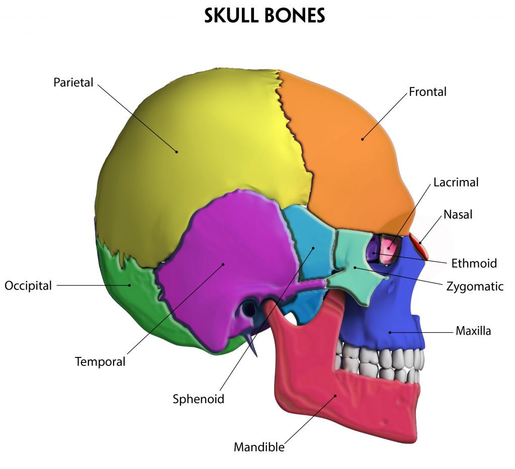
The skull is composed of several bones that protect the brain and form the framework of the face. Here are some notes about the bones of the skull:
-
- Cranial Bones:
-
- Frontal Bone: The frontal bone forms the forehead and the upper part of the eye sockets (orbits).
-
- Parietal Bones: The parietal bones are a pair of bones that form the sides and roof of the cranial cavity.
-
- Temporal Bones: The temporal bones are located on the sides and base of the skull. They contain the structures of the inner ear and house the temporomandibular joint (TMJ).
-
- Occipital Bone: The occipital bone is situated at the posterior aspect of the skull. It forms the base and back of the head and contains the foramen magnum, through which the spinal cord passes.
-
- Sphenoid Bone: The sphenoid bone is a complex bone located in the middle of the skull. It helps form the floor of the cranium, the sides of the eye sockets, and houses the pituitary gland.
-
- Ethmoid Bone: The ethmoid bone is a delicate bone located in the anterior part of the skull, between the eye sockets. It forms part of the nasal cavity and contributes to the orbital walls.
-
- Cranial Bones:
-
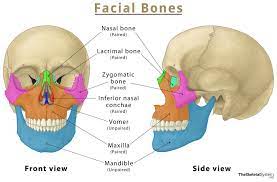
- Facial Bones:
-
- Maxilla: The maxilla is the upper jawbone and forms the central portion of the face, including the upper dental arch and the floor of the orbits.
-
- Mandible: The mandible is the lower jawbone, which forms the lower part of the face and holds the lower teeth.
-
- Zygomatic Bones: The zygomatic bones, also known as the cheekbones, form the prominences of the cheeks and contribute to the lower rim of the eye sockets.
-
- Nasal Bones: The nasal bones are small bones that form the bridge of the nose.
-
- Lacrimal Bones: The lacrimal bones are the smallest bones in the face and contribute to the medial walls of the eye sockets.
-
- Palatine Bones: The palatine bones form the posterior portion of the hard palate (roof of the mouth) and contribute to the walls of the nasal cavity.
-
- Vomer: The vomer is a thin, flat bone located in the nasal cavity. It helps divide the nasal passages.
-
- Inferior Nasal Conchae: The inferior nasal conchae are curved bones that protrude into the nasal cavity, helping to filter and humidify the air.
-
- Facial Bones:
-
- Sutures: Sutures are the fibrous joints between cranial bones, which allow slight movement during infancy and serve as areas of bone growth. Notable sutures include the coronal, sagittal, lambdoid, and squamous sutures.
Understanding the anatomy of the skull is crucial in various medical disciplines, including neurosurgery, dentistry, and forensic anthropology, as it aids in diagnosing and treating skull injuries, craniofacial abnormalities, and analyzing skeletal remains.
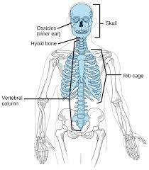
Axial Skeleton:
-
- Skull:
-
- Cranium: The cranium consists of eight bones that enclose and protect the brain.
-
- Facial Bones: The facial bones include the maxilla, mandible, zygomatic bones, nasal bones, and others, which form the structure of the face.
-
- Skull:
-
- Vertebral Column:
-
- Cervical Vertebrae: There are seven cervical vertebrae in the neck region.
-
- Thoracic Vertebrae: The thoracic vertebrae (12 in number) are located in the chest area and articulate with the ribs.
-
- Lumbar Vertebrae: The five lumbar vertebrae are situated in the lower back.
-
- Sacrum: The sacrum is a triangular bone formed by the fusion of five vertebrae.
-
- Coccyx: The coccyx, also known as the tailbone, is formed by the fusion of four coccygeal vertebrae.
-
- Vertebral Column:
-
- Rib Cage:
-
- Ribs: There are twelve pairs of ribs, which attach to the thoracic vertebrae at the back and the sternum (breastbone) at the front.
-
- Sternum: The sternum is a flat bone located in the center of the chest, consisting of the manubrium, body, and xiphoid process.
-
- Rib Cage:
-
- Hyoid Bone: The hyoid bone is a U-shaped bone located in the neck, suspended above the larynx, and not directly attached to any other bone. It plays a role in supporting the tongue and aiding in swallowing.
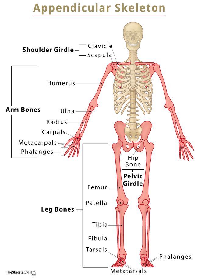
Appendicular Skeleton:
-
- Pectoral Girdle:
-
- Scapula: The scapula, or shoulder blade, is a flat triangular bone that articulates with the clavicle and humerus.
-
- Clavicle: The clavicle, or collarbone, is an S-shaped bone that connects the sternum to the scapula.
-
- Pectoral Girdle:
-
- Upper Limbs:
-
- Humerus: The humerus is the bone of the upper arm, extending from the shoulder to the elbow.
-
- Radius and Ulna: The radius and ulna are the bones of the forearm, running parallel to each other.
-
- Carpals: There are eight carpal bones in the wrist.
-
- Metacarpals: The metacarpals are five long bones that form the palm of the hand.
-
- Phalanges: The phalanges are the bones of the fingers, consisting of proximal, middle, and distal phalanges.
-
- Upper Limbs:
-
- Pelvic Girdle:
-
- Hip Bones: The hip bones, also known as coxal bones or innominate bones, consist of the ilium, ischium, and pubis. They fuse to form the pelvis.
-
- Sacrum: The fused sacral vertebrae articulate with the hip bones.
-
- Pelvic Girdle:
-
- Lower Limbs:
-
- Femur: The femur is the largest and strongest bone in the body, extending from the hip to the knee.
-
- Patella: The patella, or kneecap, is a small bone located in front of the knee joint.
-
- Tibia and Fibula: The tibia (shinbone) and fibula are the bones of the lower leg, with the tibia being larger and bearing most of the body’s weight.
-
- Tarsals: There are seven tarsal bones in the ankle.
-
- Metatarsals: The metatarsals are five long bones that form the sole of the foot.
-
- Phalanges: The phalanges in the feet are similar to those in the hands, consisting of proximal, middle, and distal phalanges.
-
- Lower Limbs:
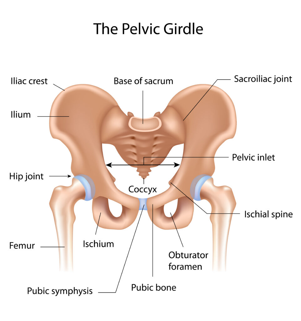
The symphysis pubis, also known as the pubic symphysis, is a specialized joint located at the midline of the pelvis.
Function: The symphysis pubis plays an important role in stabilizing the pelvis and transmitting forces between the lower limbs and the axial skeleton. It acts as a shock absorber during activities such as walking, running, and jumping.