GDA Nursing Class Note 09
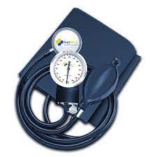
Sphygmomanometer
The sphygmomanometer is an instrument which gives an indirect
measurement of blood pressure. It consists of a gauge (manometer) to
register pressure, an inflatable compression bag with an unyielding cloth
cover (cuff), an inflating device (inflation bulb or pump), and a means of
controlled exhaust.
Basically, there are three types of gauges used in
sphygmomanometers; the mercury gravity manometer, the aneroid
manometer, and the electronic manometer.
Procedure:
Exercise: Taking Blood Pressure
1. Have the subject lying supine or seated comfortably and relaxes with the
forearm on a smooth surface at heart level.
2. The mercury meniscus or the needle of the manometer should
be at zero with the cuff deflated and the exhaust valve open.
3. Locate the brachial artery with your fingers just as you do for taking the
pulse. The brachial artery is located on the inside of the upper arm just
above the bend in the elbow on the body side.
4. The deflated cuff should be wrapped snugly (but not tightly) around the
upper arm 2 to 3 cm above the bend in the elbow. Be sure to position the
inflatable bag so about half of it is on each side of the brachial artery. When using an electronic sphygmomanometer or a cuff with a built in stethoscope
head, be sure the microphone pick up or stethoscope head is directly over the brachial artery.
5. While feeling the radial pulse in the wrist with one hand, close the
exhaust valve and rapidly inflate the cuff with the other hand. Inflate to a
pressure about 30 mm Hg above the point where the radial pulse ceases.
6. Without undue pressure, place the stethoscope over the brachial artery
just below the cuff. Exhaust the cuff at a rate of 2 to 5 mm Hg per second.
As the cuff exhausts, listen for the first clear tapping sound; the manometer
reading at this point is the systolic pressure. Continue to listen until the
sound disappears; this is the diastolic pressure.
7. Before taking the blood pressure again, allow the subject to relax for 30
to 60 seconds with the cuff deflated. Never leave the cuff inflated on the
subject for more than a few seconds.
Glucometers:
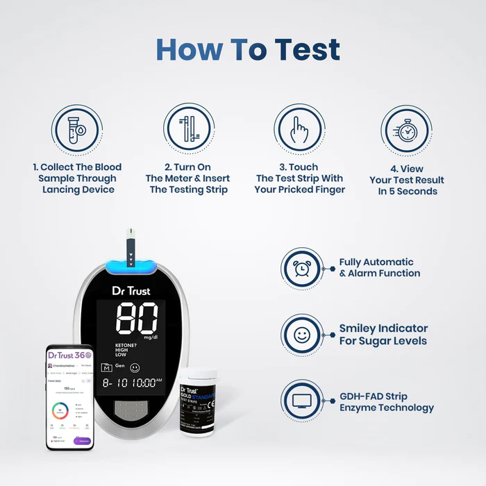
- Purpose: Glucometers are primarily used to monitor blood glucose levels, which helps people with diabetes manage their condition effectively. By tracking their glucose levels, individuals can make informed decisions about medication, diet, and lifestyle choices to keep their blood sugar within a healthy range.
- Testing Procedure: The testing process involves pricking the skin to obtain a small blood sample, typically from a fingertip. The glucometer analyzes the blood sample and displays the glucose concentration on a digital screen.
- Accuracy: Modern glucometers are generally quite accurate, but it’s essential to follow the manufacturer’s instructions carefully to ensure reliable results. Factors such as proper calibration and regular maintenance play a role in maintaining accuracy.
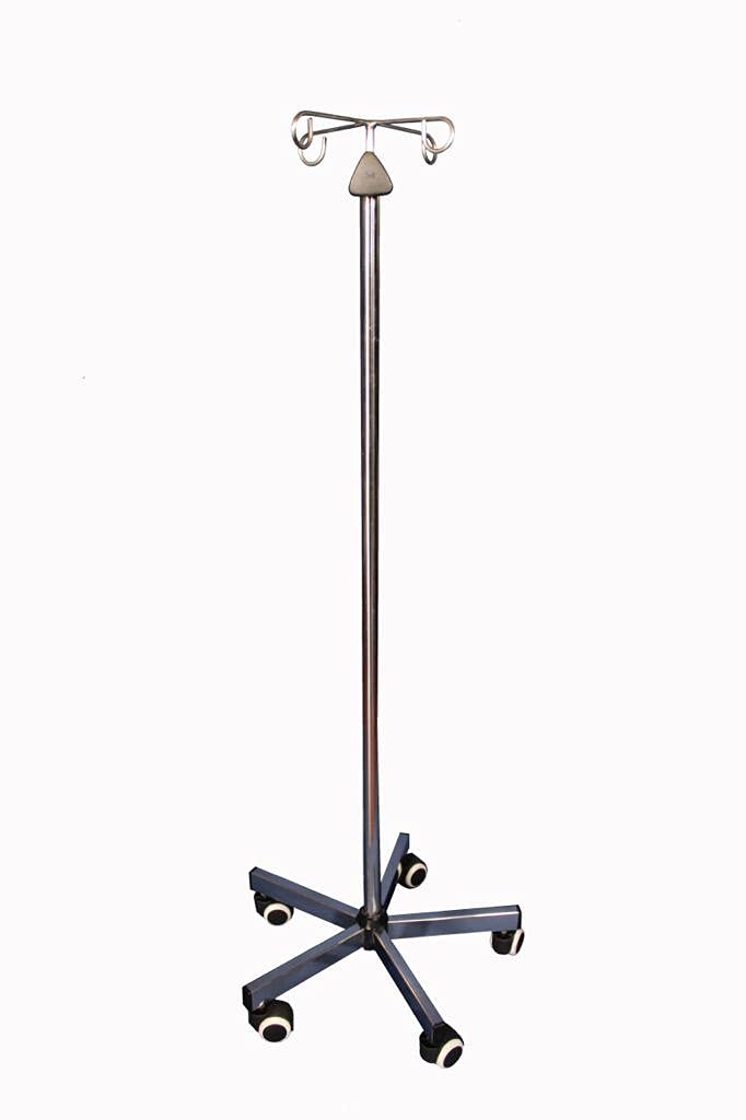
IV stands:
- Purpose: IV stands are essential tools in hospitals, clinics, and other medical facilities. They help medical professionals deliver fluids and medications safely and efficiently to patients who require intravenous therapy.
- Structure: IV stands are typically made of lightweight materials such as aluminum or stainless steel, which makes them easy to move and adjust as needed. The stand is usually equipped with a sturdy base and multiple hooks or hangers to hold IV bags, pumps, and other accessories.
- Height adjustability: Most IV stands are designed with telescoping or adjustable height features to accommodate patients of different heights or to adjust the height of the IV fluid container to control the flow rate accurately.
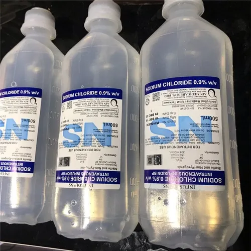
IV Fluids:
Indications for IV Fluids: a. Dehydration: Commonly caused by vomiting, diarrhea, excessive sweating, or insufficient fluid intake. b. Electrolyte imbalances: To correct low or high levels of specific electrolytes like sodium, potassium, or calcium. c. Blood loss: To replace lost volume due to bleeding. d. Surgical procedures: To maintain fluid balance during and after surgery. e. Severe infections: To support circulatory function and organ perfusion in critically ill patients.
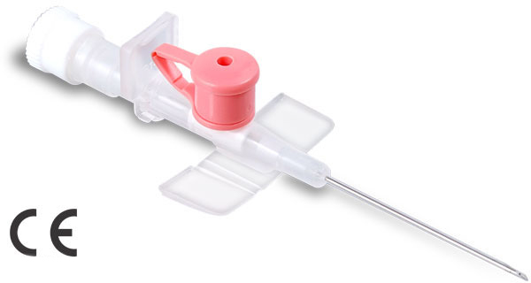
cannula:
- Definition: A cannula is a thin, hollow tube designed for insertion into the body to deliver or withdraw fluids, medications, or to provide access for medical procedures.
- Types of Cannulas: a. IV Cannula (Intravenous Cannula): Used to insert into a vein to administer IV fluids, medications, or to draw blood for diagnostic purposes. b. Nasal Cannula: A two-pronged tube used to deliver oxygen to patients through their nostrils. c. Nasogastric (NG) Cannula: Inserted through the nose into the stomach to deliver nutrients, medications, or remove stomach contents. d. Tracheostomy Cannula: Placed into the tracheostomy stoma (surgical opening in the trachea) to maintain a patient’s airway and facilitate breathing. e. Cardiac Cannula: Used during cardiac surgery to establish connections with the heart and bypass blood flow during certain procedures.
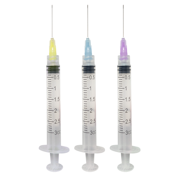
Syringe:
- Definition: A syringe is a medical device used for injecting or withdrawing fluids from the body. It consists of a cylindrical barrel with graduated markings, a plunger, and a tip (needle or nozzle) for delivering or extracting substances.
- Types of Syringes: a. Standard Syringe: The most common type, with a needle attached to the tip for injections or blood draws. b. Insulin Syringe: Specifically designed for administering insulin to diabetic patients, with fine and short needles for subcutaneous injections. c. Tuberculin Syringe: Used for administering small volumes of medication, such as TB tests or allergy injections, with a long and thin needle. d. Oral Syringe: A type of syringe without a needle, used to administer liquid medications orally.
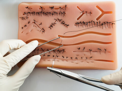
Suture:
- Types of Sutures: Sutures come in various materials, each with specific properties. Common types include:
- Absorbable sutures: These sutures break down over time and are gradually absorbed by the body. They are typically used for internal tissues and eliminate the need for suture removal.
- Non-absorbable sutures: These sutures do not break down and require manual removal after the wound has healed. They are often used for external closures and in situations where long-term wound support is needed.
- Suture Materials: Suture materials can be made from natural sources (e.g., silk or catgut) or synthetic materials (e.g., nylon, polypropylene, or polyester). The choice of material depends on the type of tissue being sutured, the location of the wound, and the expected healing time.
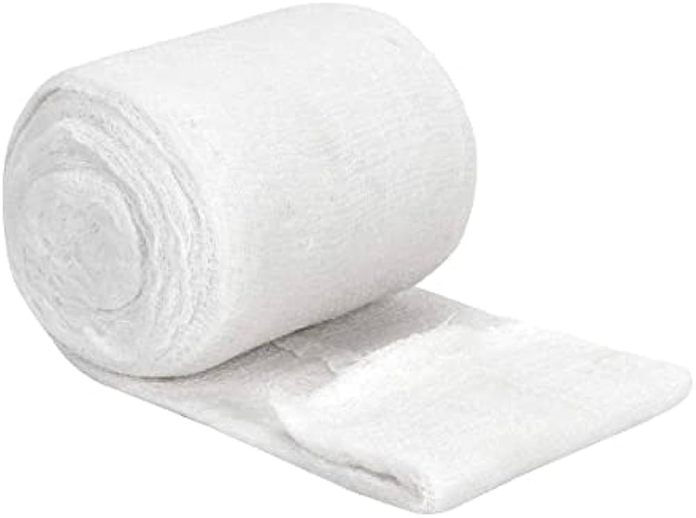
Gamjee:
- Definition: Gamjee is a type of cotton-based surgical dressing that is used to cover wounds and injuries.
- Composition: Gamjee dressings typically consist of several layers. The innermost layer is made of cotton wool, which provides cushioning and absorbs exudate (fluids from the wound). The outer layers are made of gauze or other fabric materials that hold the cotton wool in place and keep the dressing secure.
- Medication: Gamjee dressings are often medicated with antiseptic or antibacterial substances. The medication helps prevent infection and promotes wound healing.
intruments for dressing tray:
- Scissors: Different types of scissors may be present, including bandage scissors, Mayo scissors (used for cutting tough tissues), and fine-point scissors for delicate work.
- Forceps: These are used to grasp and hold tissues, dressings, or other objects during procedures. Examples include tissue forceps, Adson forceps, and dressing forceps.
- Hemostats: Also known as arterial forceps, they are used to clamp blood vessels and control bleeding during surgical procedures.
- Needle holder: Used to hold and manipulate surgical needles during suturing.
- Scalpel: A surgical knife used for making incisions in the skin or tissues.
Needle holders:
- Handle Design: The handles of a needle holder are designed for a comfortable grip and precise control. They may feature a ratchet mechanism, which allows the surgeon to lock the jaws in a closed position to securely hold the needle during suturing.
- Jaw Design: The jaws of a needle holder have a serrated or crosshatched surface, which provides a firm grip on the surgical needle, preventing it from slipping while suturing.
- Types of Needle Holders: There are various types of needle holders available, including Mayo-Hegar, Olsen-Hegar, Webster, and Castroviejo needle holders. The main difference lies in the combination of needle holding function and additional features like scissors in some models.